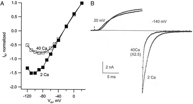
Figure 1.
Block of Shaker K channels by external Ca2+ at negative membrane voltage. (A) The curves give the current through a set of open channels in 2 and 40 mM Ca2+ as a function of membrane voltage (Vm). The channels were activated by a first 5-ms step to 40 mV, and a test step was then applied, with successive trials for the test step in the range of 20 to −120 mV. Current was measured just after the beginning of the test step, before the gates could respond to the new voltage (most gates open). Current for each solution is normalized relative to its value at 20 mV (the current at 20 mV was almost identical in the two solutions). External solutions: 2 or 40 Ca2+ with 40 K+. (B) The traces show IK of Shaker-IR channels during an activating step to 20 mV followed by a step to −140 mV, in 40 and 2 mM Ca2+. There is an obvious fast phase of decay in 40 Ca2+ caused by Ca2+ block. Solutions: 40 Ca 25 K or 2 Ca 40 K//150 K.

