
Figure 4.
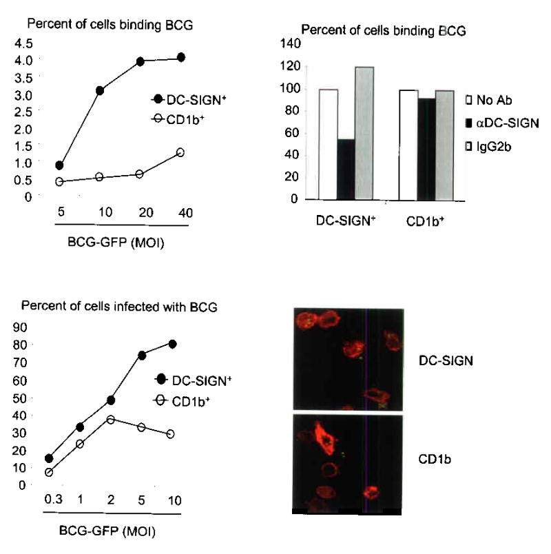
DC-SIGN+ macrophages bind and phagocytose mycobacteria. (a) Cytokine-differentiated monocytes were cultured with M. bovis BCG. Data represent percent of cells that are double positive for either DC-SIGN and BCG-GFP (filled circle) or CD1b and BCG-GFP (open circle) from two independent experiments. (b) Binding was measured in the presence of a DC-SIGN blocking antibody or controls. Data are represented as percent of binding relative to media control. (c) DC-SIGN+ and CD1b+ cells were incubated with BCG-GFP and uptake was measured by double labeling. Data is representative from two independent experiments. (d) Confocal images of cells cultured and labeled as in (c).