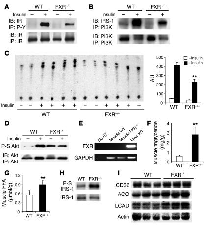
Figure 4. Impaired insulin signaling and upregulation of genes involved in fatty acid metabolism in muscle of FXR–/– mice.
Muscle tissue homogenate from 4–5 mice per group were pooled together and subjected to IP and IB using antibodies as indicated. Northern blot analysis was performed on individual mice. Results are representative of at least 3 independent experiments. (A) Phosphorylation of IR after insulin stimulation (1 U/kg). Muscle homogenates were subjected to IP by anti-phosphotyrosine (P-Y) antibody 4G10 and IB by IR antibody. Total IR level was analyzed by IP followed by IB using the IR antibody. Quantitation was derived from 3 independent experiments. (B) Level of PI3K-associated IR after insulin stimulation as analyzed by IP using PI3K antibody followed by IRS-1 IB. (C) PI3K activity assay using immunopricipitates by anti-phosphotyrosine antibody. Muscle homogenates from individual mice were subjected to IP by anti-phosphotyrosine antibody followed by PI3K assay. Quantitation was derived from individual mice. (D) Phosphorylation of Akt (serine 473; P-S Akt) after insulin stimulation. (E) FXR expression by RT-PCR. (F and G) Analysis of intramuscular triglyceride and FFA content (n = 8 per group). (H) Serine 307 phosphorylation (P-S) of IRS-1 in the muscle after IP using IRS-1 antibody. (I) Expression of genes involved in fatty acid transport and oxidation in WT and FXR–/– muscle. ACO, acyl-CoA oxidase; LCAD, long-chain acyl-CoA dehydrogenase. **P < 0.01.

