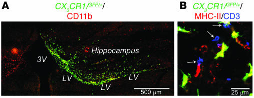
Figure 8. Distribution of CX3 CR1/GFP/+ MG 9 days after stereotaxic injection.
(A) Localization of GFP+ cells colabeled with the microglial marker CD11b in the right lateral ventricle (LV) after injection of MG(IL-4) from CX3CR1/GFP/+ mice into mice with EAE. Note the hippocampal area in the MG(IL-4)-injected mice was heavily populated by MG(IL-4)-GFP+ cells. (B) MG(IL-4)-GFP+ cells coexpressing MHC class II populated the spinal cord at level T8–T9 adjacent to the ependyma of the central canal associated with CD3+ cells (arrows). 3V, third ventricle.

