
2.
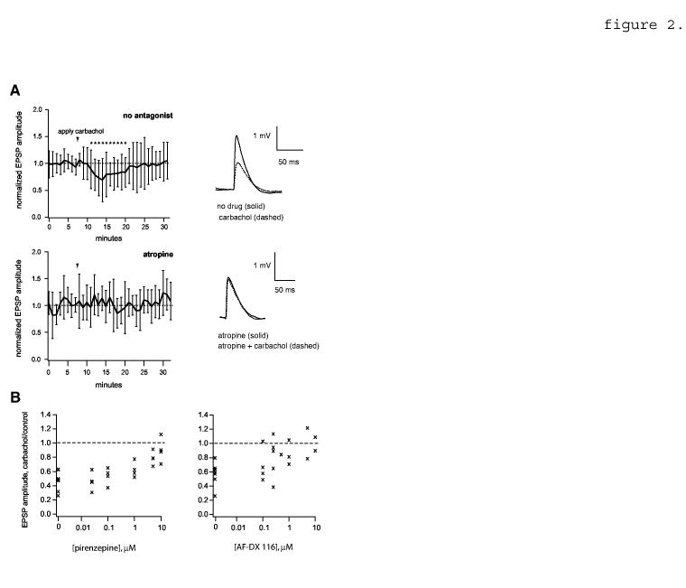
EPSP reduction by carbachol; effect of muscarinic receptor antagonists. A, Top left, EPSP amplitude time course showing the effect of carbachol in the absence of 1 μM atropine (n=20 pairs). EPSPs are averaged in 1 minute bins and normalized to the mean value before carbachol application. Arrowhead shows the time of initial carbachol application (duration varied between 2 and 3 min). Asterisks indicate time points that differ significantly from control (p<0.05, two-tailed unpaired t-tests). Top right, example average traces showing the effect of carbachol on an example pair (c.f. figure 1A). Resting Vm = -64 mV. Bottom left, data obtained in the presence of 1 μM atropine (n=6 pairs). Bottom right, example traces; resting Vm = -61 mV. B, antagonism of carbachol-mediated EPSP suppression by the M1-subtype specific antagonist pirenzepine (left) and the M2 subtype-specific antagonist AF-DX 116 (right). Each point represents average peak amplitude data from one pair at the given antagonist concentration (n=7 pairs total for pirenzepine and 8 pairs for AF-DX 116). Pirenzepine was tested at 0.025, 0.1, 1, 5 and 10 μM and AF-DX 116 at 0.1, 0.25, 0.5, 1, 5 and 10 μM.