3.

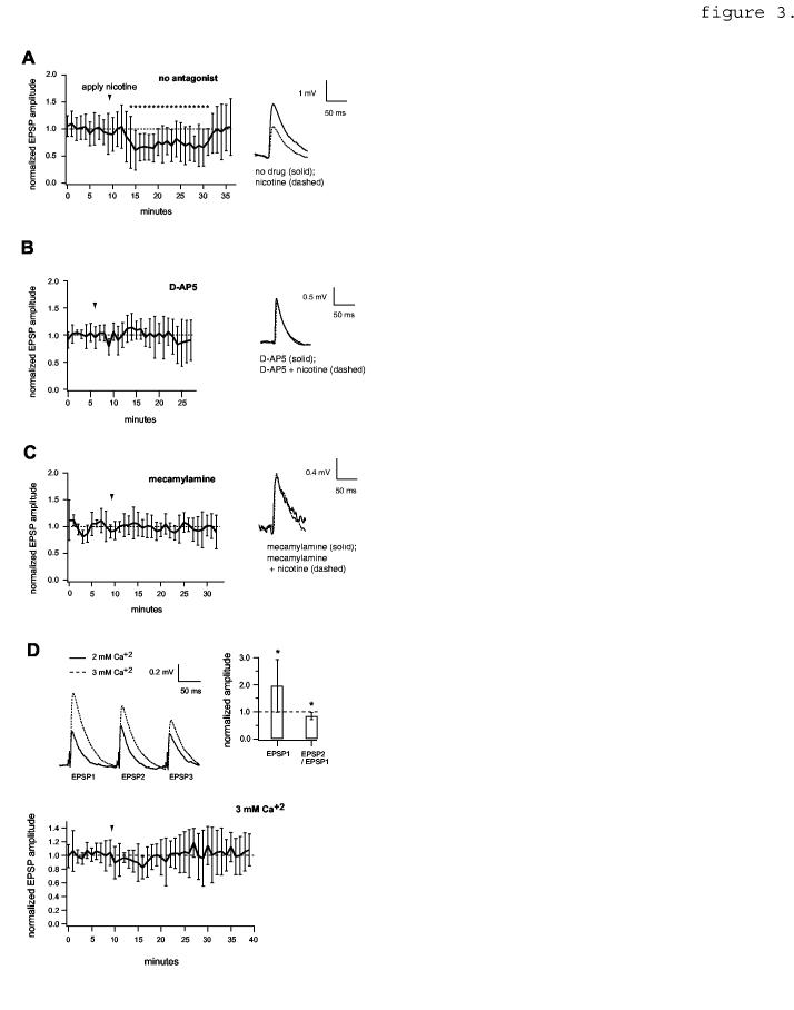
NMDA receptor dependence of EPSP reduction by nicotine. A, Left, EPSP amplitude time course showing the effect of nicotine in the absence of antagonist, normalized to the value preceding nicotine (n=20 pairs). Asterisks indicate time points that differ significantly from control (p< 0.05, two-tailed unpaired t-tests). Right, average traces showing the effect of nicotine on an example pair (c.f. figure 1E). Resting Vm = -67 mV. B, data obtained in the presence of 50 μM D-AP5 (n=6). For average EPSP traces (right), resting Vm = -62 mV. C, data obtained in the presence of the selective nicotinic receptor antagonist, 10 μM mecamylamine (n=7 pairs). Resting Vm = -66 mV. Note: apparent background noise seen in this example was atypical. Data in A,B,C were obtained in 0 mM Mg+2. D, Top, [Ca+2]-dependent changes in EPSP amplitude and paired-pulse ratio. Asterisks denote p< 0.05 (n=9 pairs). Bottom, effect of nicotine in ACSF containing 1 mM Mg+2 / 3 mM Ca+2 (n=6 pairs).
