6.

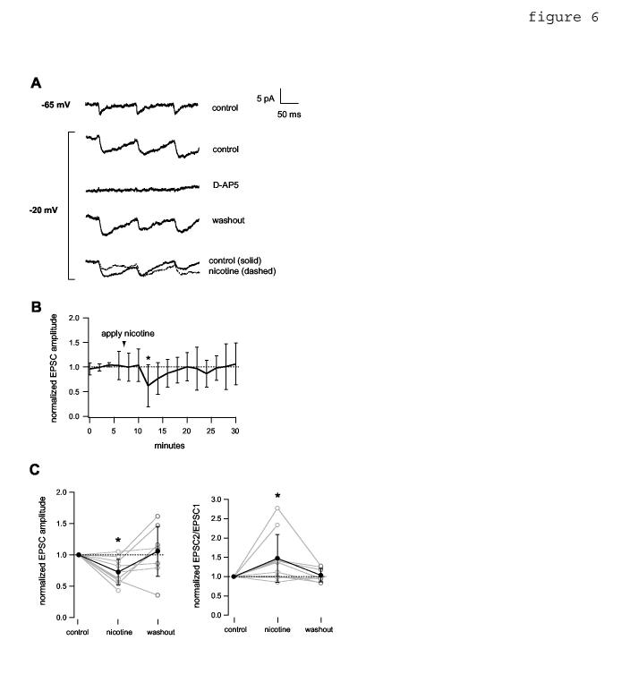
Depolarization of the postsynaptic neuron in 1 mM Mg+2 ACSF permits nicotinic reduction of NMDAR-mediated EPSCs. Data were obtained in the presence of 10 μM DNQX. The soma of the postsynaptic cell was held in voltage clamp at the indicated holding potential. A, average current traces showing NMDAR-mediated EPSCs evoked by 10 Hz presynaptic stimulation. Depolarization to -20 mV enhanced the NMDAR current (compare top 2 traces). The evoked EPSC was blocked reversibly by bath-applied 50 μM D-AP5. Bottom traces, effect of bath-applied (3 min) 10 μM nicotine. B, average EPSC amplitude time course, normalized to the mean value before nicotine application (n= 7 pairs). Asterisk indicates time point that differs significantly from control (p< 0.05, two-tailed unpaired t-test). C, Left, normalized EPSC amplitude data for all pairs. Mean values (filled circles) were 0.73 +/- 0.20 during nicotine and 1.05 +/- 0.40 after washout. Asterisk denotes significance relative to control (p< 0.01, n= 10 pairs). Right, normalized EPSC2/EPSC1 for all pairs. Mean values (filled circles) were 1.47 +/- 0.62 during nicotine and 1.04 +/- 0.17 after washout. Asterisk denotes significance relative to control (p< 0.05, n= 10 pairs). Connections where mean control EPSC peak amplitude was < 2.5 pA were excluded because of poor signal:noise ratio. Washout data were not obtained for two pairs.
