
Fig. 2.
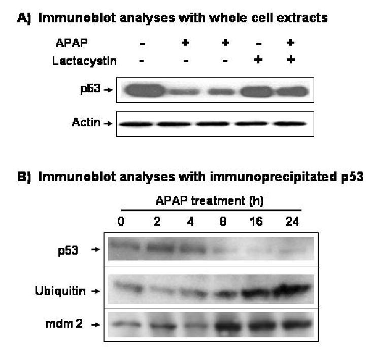
Time- and ubiquitin-dependent degradation of p53 after exposure to APAP. (A) Effect of lactacystin on APAP-induced p53 degradation. C6 cells were pretreated with 1 μM lactacystin for 4 h before cells were exposed to APAP for additional 24 h in the presence of 1 μM lactacystin. The level of p53 in the soluble fractions (100 μg protein/well) was then determined by immunoblot analysis with the specific antibody against p53 (top) or actin (bottom). (B) Immunoblot analyses of immunoprecipitated p53. Immunoprecipitation of p53 was performed as described under Materials and Methods. The immunoprecipitated p53 protein was washed twice with 1 x PBS and subjected to 10% SDS-PAGE followed by immunoblot analysis with the respective antibody against p53 (top), ubiquitin (middle), or mdm2 (bottom). These results represent a similar pattern of three independent experiments of p53 immunoprecipitation.