
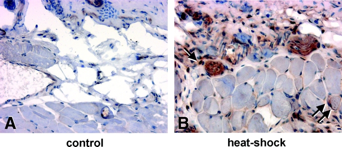
FIGURE 1. Longitudinal sections of flap tissue 24 hours after preconditioning stained for heat shock protein (HSP)-32 expression in control animals (A) and animals after local heat shock exposure (B). Note the nuclear and cytosolic staining of heat shock-treated flaps within the subcutaneous (arrow) and muscle tissue (double arrow) (original magnification ×80).