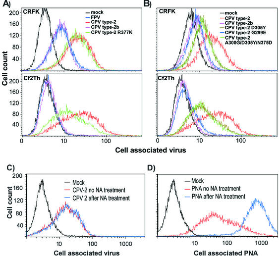
FIG. 3.
(A) Binding and uptake of FPV (blue), CPV type 2 (red), CPV type 2b (purple), or CPV type 2-R377K (green) capsids incubated with feline CRFK cells or canine Cf2Th cells. Cells were incubated with 20 μg of capsid/ml for 1 h at 37°C and then detached with EDTA, fixed, and permeabilized; the cell-associated virus was quantified with a Cy2-labeled anti-capsid monoclonal antibody, followed by analysis by flow cytometry. (B) Binding and uptake of CPV type 2 (red), CPV type 2b (purple), CPV type 2 D305Y (dark green), CPV type 2 G299E (blue), and CPV type 2 A300G/D305Y/N375D (light green) capsids into CRFK or Cf2Th cells as described above. (C and D) Binding of CPV type 2 capsids to CRFK cells that were either mock treated (red) or incubated with neuraminidase (blue). Cells were incubated with CPV type 2 (C) or fluorescein isothiocyanate-labeled peanut agglutinin (PNA) (D).

