FIG. 5.
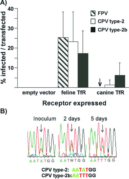
(A) Virus susceptibility of TRVb cells expressing feline or canine TfRs. Cells transfected with plasmids expressing the feline or canine TfRs were inoculated with CPV type 2, FPV, or CPV type 2b and then incubated for 24 h before incubation for 30 min with Texas Red-labeled canine Tf. After fixation and permeabilization virus infection detected by staining for the viral NS1 protein. The bars represent one standard deviation of the mean of the percentage of Tf-binding cells that became infected in six separate experiments. (B) The replacement of CPV type 2 by CPV type 2a when the two viruses were grown together in TRVb cells expressing the canine TfR for 5 days. The inoculum contained nine times more TCID50 of CPV type 2 than CPV type 2b, which was reflected in the double sequencing profile at position 3046. Samples of virus were collected from the culture inoculum and from the culture at days 2 and 5 after inoculation. The viral DNA was amplified by PCR and sequenced, and the profiles of sequences from nucleotides 3043 to 3049 are shown.

