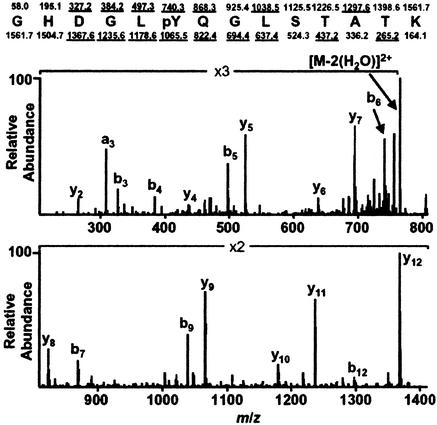
Figure 1.
MS/MS spectrum of the CD3ζ-derived peptide GHDGLpYQGLSTATK. Predicted nominal masses of type b and type y ions appear above and below the sequence, respectively. Those observed are underlined. Note that the mass difference between y8 and y9, and also b5 and b6, is 243 Da, corresponding to a phosphotyrosine residue.

