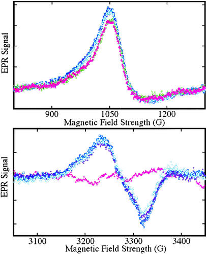
Figure 6.
EPR spectra from Fe(II)NO and Fe(III) hemes obtained by exposure of hemoglobin to nitrite. EPR spectra were obtained at 9.28 GHz, with 10 mW incident power and field modulation of 20 G at 100 kHz. The external field was scanned for 4 min with a detection time constant of 0.128 sec over to range: 800−1,300 G to detect high-spin heme-Fe(III) (Upper) or 3,050−3,450 G to detect heme-Fe(II)NO (Lower). A spectrum of a sample of a neat 3.8 mM oxyhemoglobin solution, frozen in liquid nitrogen, is displayed (▴, pink) in both panels; the trace (Lower) was used as a base-line for subtraction from subsequent scans. Spectra were subsequently obtained from samples withdrawn from the solution after incubation with 37 μM nitrite for periods of 5 (□, blue), 12 (⧫, purple), and 24 (○, teal) min, followed by deoxygenation and freezing in liquid nitrogen. SNO-Hb is observed on reoxygenation of such samples. A spectrum obtained without deoxygenation after an incubation time of 10 min (◊, green) is also shown (Upper).

