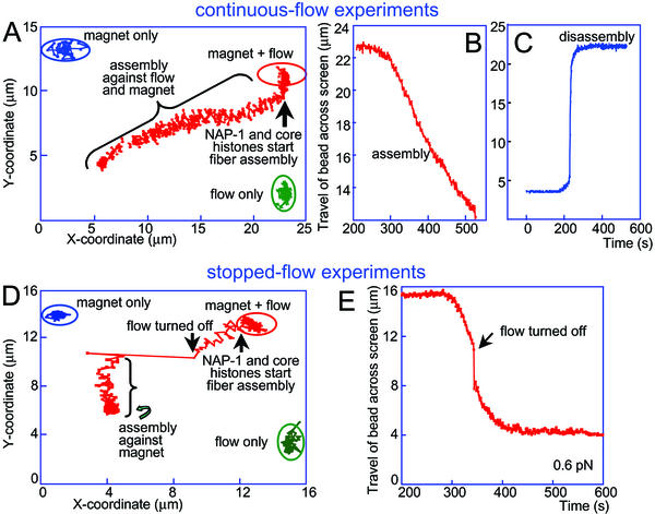
Figure 3.
Bead coordinates in a continuous-flow experiment (A) and a stopped-flow experiment (D). The positions of the bead at the beginning of the experiment (magnet only), at the point where flow was initiated (magnet + flow), and a control detection of the bead position when only flow was applied (flow only) are encircled. The entrance of core histones and NAP-1 into the cuvette led to initiation of the assembly process (movement of the bead leftward and downward). The jump accompanying the stopping of the flow in D was followed by assembly under force exerted on the bead by the external magnet only. (B and E) Assembly curves (distance traveled by bead across the screen as a function of time) in a continuous-flow (B) and in a stopped-flow (E) experiment. (C) NaCl (2 M) was flowed into the cell immediately after completion of assembly to observe the disassembly process. The DNA molecule in the continuous-flow experiment (A–C) was presumably an end-to-end dimer of the λ-DNA monomeric genome. Such dimers can occasionally occur as a result of hybridization of the λ-DNA cos sites during the end-labeling procedure.

