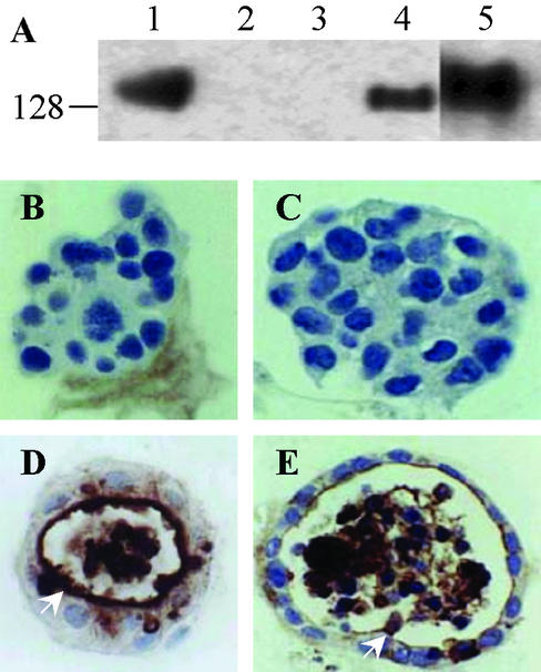
Figure 2.
CEACAM1-4S mediated reversion of MCF7 cells to normal phenotype. (A) Cell lysate preparations (60 μg of total protein) from cells grown on plastic were separated by 8% reducing SDS/PAGE gel and immunoblotted with anti-CEACAM1 mAb (4D1C2). Lane 1, positive control HT29 colon carcinoma cells; lane 2, parental MCF7 cells; lane 3, MCF7/pHβ (vector); lane 4, MCF7/CEACAM1-4S; lane 5, MCF7/CEACAM1-4L. (B–E) Cells (2.5 × 10⁵ per well) were grown in Matrigel for 12 days, fixed, and embedded in paraffin, and sections were stained with anti-CEACAM1 mAb (4D1C2). (B) MCF7 cells exhibiting no lumen formation. (C) Vector-transfected MCF7 cells exhibiting no lumen formation. (D) Nonmalignant mammary epithelial cells MCF10F exhibiting lumen formation. (E) MCF7/CEACAM1-4S-transfected cells exhibiting lumen formation. (Magnification, ×600.)

