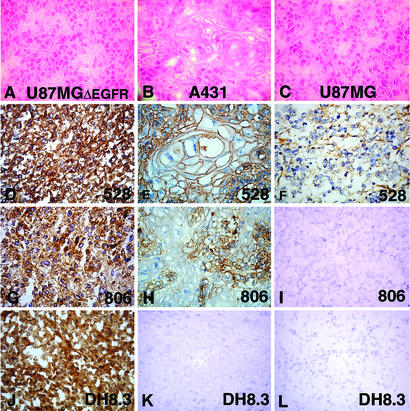
Figure 1.
Hematoxylin/eosin (A–C) and immunohistochemical (D–O) (avidin–biotin-complex technique, diaminobenzidine tetrahydrochloride chromogen) staining of tumor xenografts U87MGΔEGFR (A, D, G, and J), squamous cell carcinoma cell line A431 (B, E, H, and K), and parental (nontransfected) U87MG cell line (C, F, I, and L) with 528 (D–F), 806 (G–I), DH8.3 (J–L). (D, G, and J) Intense staining of U87MGΔEGFR with all three antibodies. (E) Intense staining of A431 with 528. (H) Less intense staining of A431 with 806. (K) Negative staining of A431 with DH8.3. (F) Weak focal staining of parental U87MG with 528. (I and L) No staining of parental U87MG with 806 or DH8.3.

