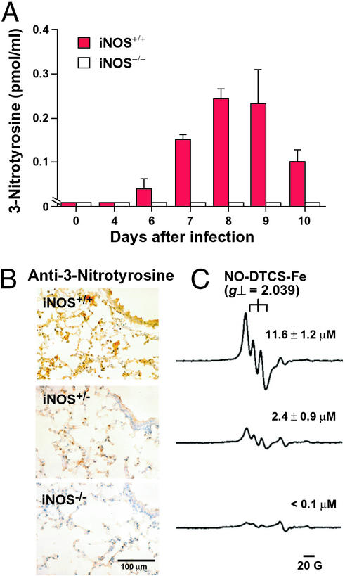
Figure 4.
3-Nitrotyrosine and NO formation in influenza virus-infected lungs from wild-type and iNOS-deficient mice. (A) HPLC/electrochemical-detection analysis of protein in BAL fluid was used to determine tyrosine nitration. Concentrations of protein-bound 3-nitrotyrosine in BAL fluid (means ± SE, n = 3) are plotted versus time. (B) Immunohistochemistry for 3-nitrotyrosine formation in lung tissues. (C) Typical ESR signals of the NO-N-dithiocarboxy(sarcosine)-Fe2+ adduct for each corresponding group in B. The amounts of NO-N-dithiocarboxy(sarcosine)-Fe2+ as assessed by double integration of ESR spectra (15) are shown (means ± SE, n = 3) (P < 0.01 by t test for the value for iNOS+/+ vs. values for iNOS+/− and iNOS−/−).

