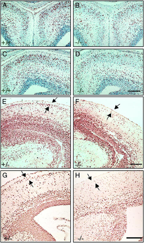
Figure 3.
Proliferation and migration of neurons in brains of ERβ−/− mice and their WT littermates. (A–D) Coronal sections (6 μm, paraffin) of the anterior telencephalon from male littermates were stained with anti-BrdUrd antibody and counterstained with hematoxylin. Cortical neurons generated at E14.5 and E15.5 and labeled with BrdUrd were analyzed at E17.5. Note fewer BrdUrd-labeled neurons in the ERβ−/− brain (B and D), especially the superficial layer of the cerebral cortex, compared with littermate control (A and C). (Scale bar, 50 μm.) Comparable coronal sections (6 μm, paraffin) from male (F) and female (H) ERβ−/− mice and their +/− littermates (E and G) are shown. Cortical neurons generated at E15.5 and E16.5 and labeled with BrdUrd were analyzed at E18.5. Note that the BrdUrd-labeled neurons in the ERβ−/− brain (F and H) are distributed diffusely in the cerebral cortex and are not organized into a well defined layer (arrows) at the superficial part in the cortex as in the control brains (E and G). (Scale bar, 50 μm.)

