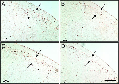
Figure 4.
Fate in the postnatal brain of neurons generated at E15.5 and E16.5. Neurons generated and labeled at E15.5 and E16.5 were analyzed at P14. Comparable coronal sections (6 μm, paraffin) from male (A and B) and female (C and D) littermates were stained with anti-BrdUrd antibody. Note the decreased numbers of BrdUrd-labeled neurons distributed in the lamina II–III of cortex (arrows) in ERβ−/− brains (B and D) compared with littermate controls (A and C). (Scale bar, 50 μm.)

