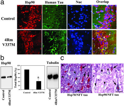
Figure 1.
The tau and Hsp70/90 levels in hippocampus of mutant tau Tg mice and in AD brain. (a) Hippocampal sections of control and mutant tau (4Rm V337M) Tg mice were analyzed for intraneuronally accumulated tau and Hsp90 by immunofluorescence confocal microscopy. Images were overlapped with nuclear counterstaining (Nuc). White arrowheads indicate those neurons with intensive immunoreactivity for accumulated tau but little signal for Hsp90. Black arrowheads indicate neurons with strong immunostaining for Hsp90 but lacking immunoreactivity for accumulated tau. (Bar = 10 μm.) (b) Hippocampal tissues were homogenized and Hsp90 was assayed by Western blotting. Levels of β-tubulin were similar between control and Tg mice. Quantitative data represent averages of three experiments (mean ± SE); *, P < 0.01, with respect to control. (c) Intraneuronal immunostaining for NFT tau, Hsp70, and Hsp90 in hippocampal sections of an AD patient. Adjacent sections were doubly immunostained for hyperphosphorylated (NFT) tau (red, arrows) and for either Hsp70 or Hsp90 (blue, arrowheads). (Bar = 40 μm.)

