Figure 1.
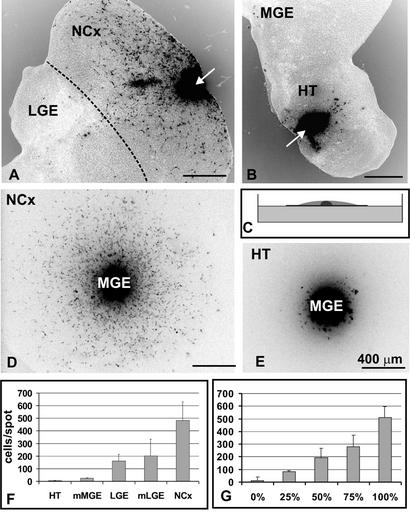
Different regions of the developing brain are differentially permissive for MGE cell migration. (A) PKH26-labeled MGE cells (black) were transplanted into neocortex in brain slice cultures (arrow). Forty-eight hours later, many MGE cells had dispersed throughout the neocortex. Note that only a few cells crossed the boundary back to the LGE (broken line). (B) MGE cells grafted into the hypothalamus remained at the graft site (arrow), unable to penetrate into the host brain tissue. (C) Diagram of the spot assay. Dissociated cells (gray) were spotted on polycarbonate membrane floating on the surface of culture medium in a petri dish, and a reaggregate of labeled MGE cells (black) was placed in the center of the spot. Cells were cultured for 48 h. (D) Labeled MGE cells (black) readily disperse through neocortical cells. (E) MGE cells do not migrate into a spot of hypothalamic cells. (F) Quantification of MGE cell migration through spots of different brain regions (number of cells per spot ± SD). (G) Migration of MGE cells through spots containing mixed neocortical and hypothalamic cells. The percentage indicates the amount of neocortical cells in a particular spot (number of cells per spot ± SD). NCx, neocortex; HT, hypothalamus; mMGE, mantle zone of the MGE; mLGE, mantle zone of the LGE. Scale bars = 400 μm.

