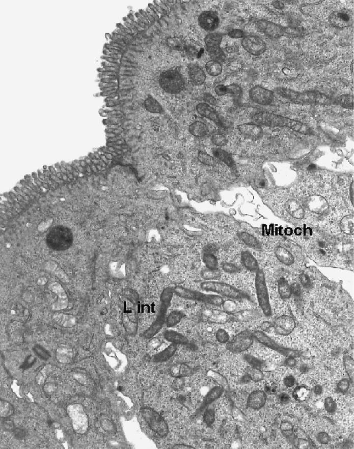
Figure 3.

Transmission electron micrograph of ultrathin section of lower villous area of porcine ileum xenograft, showing numerous intracytoplasmic L. intracellularis (L int), adjacent mitochondria (Mitoch), and mature microvilli and other organelles typical of mature intestinal epithelial cells. Magnification × 2200.
