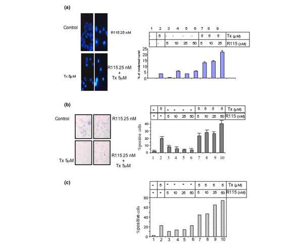
Figure 3.

Nuclear condensation of cells incubated for 5 days with tamoxifen (Tx) and R115,777. (a) Cells were fixed and stained with the DNA intercalating agent DAPI (left) and nuclei were examined by fluorescence microscopy. DAPI-stained nuclei were counted as normal or condensed nuclei (right). One randomly selected field is presented for each treatment. For three independent experiments, quantification of at least 200 nuclei was performed for each treatment. Error bars indicate the mean values ± standard error of the mean. (b) Cells were fixed and stained for the detection of the caspase cleavage product of cytokeratin 18 by immuno-histochemistry. Floating and adherent cells are represented in cytospin preparations. These preparations were fixed and stained with monoclonal antibody M30 CytoDeath. The proportion of positive cells (brown) was calculated as a percentage of the total number of cells. All determinations were performed in triplicate with at least 400 cells being counted. Error bars indicate the mean values ± standard error of the mean. (c) Cells were fixed and stained for the detection of the caspase cleavage product of cytokeratin 18 by FACS. Floating and adherent cells were harvested, fixed and stained with the fluoresein-conjugated monoclonal antibody M30 CytoDeath and analysed using flow cytometry. The proportion of positive cells was calculated as a percentage of the total number of cells. Data are representative of one to three independent experiments; intra-assay variations were <1%.
