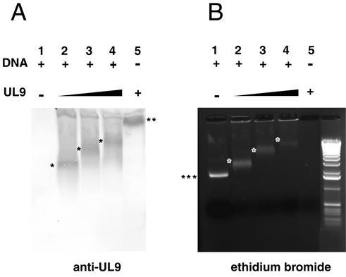
FIG. 11.
UL9/ssDNA complexes are detected by Western blotting. ssDNA-binding reaction mixtures containing 400, 800, and 1,000 nM UL9 protein (indicated by the thickness of the black triangle above the gel) with R113/F115A mutations and containing DNA (+) were resolved on a 1% agarose gel (B), electrotransferred to an Immobilon P membrane, and probed with an anti-UL9 antibody (A). The positions of free ssDNA (three asterisks) and UL9 alone (two asterisks) are indicated at the side of the gel. The positions of UL9/ssDNA complexes are indicated by the single asterisks in the gel.

