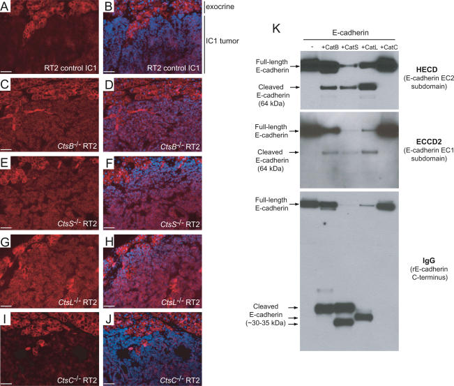
Figure 6.
E-cadherin is a target substrate for cathepsins B, L, or S, but not C, in vivo and in vitro. E-cadherin staining was used to confirm the H&E grading and revealed a consistent maintenance of E-cadherin levels even in microinvasive carcinomas (IC1) from CtsB−/− RT2 (C,D), CtsS−/− RT2 (E,F), and CtsL−/− RT2 (G,H) mice, but not in CtsC−/− RT2 (I,J) or control RT2 (A,B) IC1 tumors. E-cadherin-stained images (red) are shown in the left panels, with E-cadherin/DAPI merged images in the right panels. The normal exocrine tissue (in which levels of E-cadherin are the same in all genotypes) is shown at the top of the image for comparative reference to the IC1 tumor below. Bars, 50 μm. (K) Recombinant E-cadherin is cleaved by cathepsins B, L, and S, but not cathepsin C in vitro. E-cadherin was incubated under identical conditions with each activated cathepsin, as indicated above the top panel, and Western blots were hybridized with HECD-1 (E-cadherin extracellular subdomain EC2), ECCD2 (extracellular subdomain EC1), or IgG (to detect recombinant E-cadherin, which has an IgG linker at the C terminus). Full-length and cleaved E-cadherin fragments are indicated at the left side.

