Figure 7.
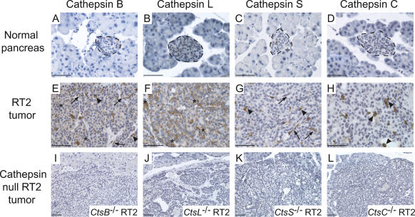
Cell-type-specific expression of cathepsins in mouse RT2 pancreatic tumors. Normal pancreas and RT2 tumors were stained with antibodies against cathepsins B, L, S, and C as indicated. (A–D) Representative images of normal mouse pancreas stained for each antibody are shown in the first row, with normal islets indicated with a dotted black line, surrounded by normal exocrine cells. (E–H) Representative images of RT2 tumors stained for each antibody are shown in the second row. Cathepsin-positive cells are stained in brown, and hematoxylin (blue) was used as a counterstain. Tumor cell staining is indicated by asterisks, endothelial cell staining by arrows, and immune cell staining by arrowheads. (I–L) The specificity of antibodies against cathepsins B, C, L, and S is demonstrated by the absence of staining in the corresponding cathepsin-null/RT2 tumors, as indicated in the third row. Bars, 50 μm.

