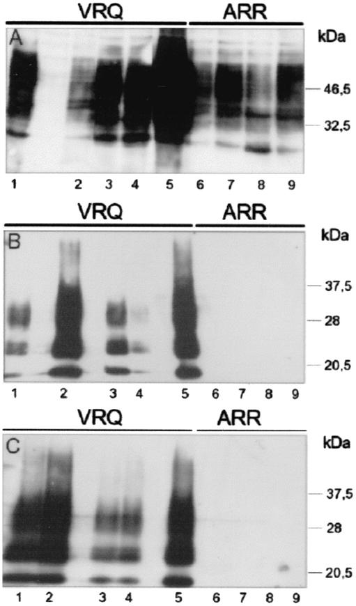
FIG. 1.
Analysis of normal and abnormal PrPs in Rov clones expressing the VRQ or the ARR allele of ovine PrP after exposition to sheep prion. Transmission of sheep prion obtained from a VRQPrP animal to nine Rov clones was tested by incubating confluent monolayers expressing the VRQ (lanes 1 to 5) or the ARR (lanes 6 to 9) PrP allele with infectious brain homogenate for 2.5 days. The inoculum was removed, and the cells were washed and passaged 2.5 days later. Cultures were then subcultured once a week at a one-fourth dilution. Parts of the cultures were lysed and analyzed for PrP at either one (A and B) or six (C) passages p.i.. Lane 1, Rov9; lane 2, RovC79; lane 3, RovA7; lane 4, RovA1; lane 5, Rov5; lane 6, RovG2; lane 7, RovE2; lane 8, RovG4; lane 9, RovE5. Some lanes were not loaded. (A) Normal PrP in the different clones at one passage p.i. Proteins (the equivalent of 25 μg) were methanol precipitated, and PrP was analyzed by immunoblotting with 4F2 MAb directed against the NH2 terminus region of PrP. Since most of the Rov-derived PrPsc undergoes a proteolytic cleavage that removes a portion of the NH2 terminus region (unpublished observation), accumulated PrPsc in Rov cells cannot be detected by 4F2 MAb. (B and C) Abnormal PrP was isolated from PK-digested cell lysates (the equivalent of 250 μg of proteins) at one (B) or six (C) passages p.i. The level of PK resistance of PrPsc generated in infected VRQ-Rov cells is high enough to sustain digestion with high amounts (50 μg/ml) of PK (28). However, in these experiments, low concentrations of PK (ca. 5 μg/ml) were used to detect any abnormal PrP with possible low levels of PK resistance. Immunoblots were stained with 2D6 MAb raised against the COOH terminus region of PrP.

