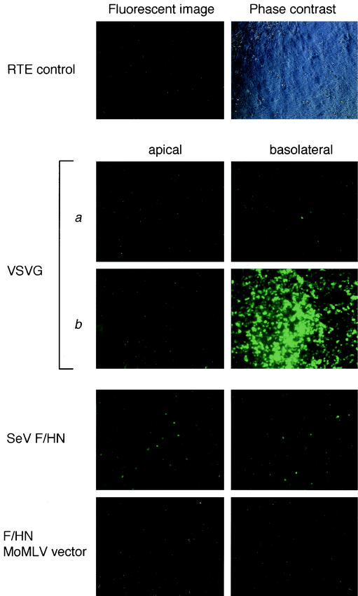
FIG.7.
Transduction of RTE cells with pseudotyped SIVagm vectors. Fluorescent images of EGFP expression on day 3 and phase contrast microscopy of RTE cells. RTE cells were exposed to Fct4- and SIVct+HN-pseudotyped SIVagm vectors at the apical or basolateral side. The titers of the VSV-G-pseudotyped SIVagm vector were (a) 4.9 × 10⁵ T.U./0.1 ml and (b) 1.2 × 10⁸ T.U./0.1 ml; the titer of the SeV Fct4- and SIVct+HN-pseudotyped SIVagm vector was 1.2 × 10⁴ T.U./0.1 ml; and the titer of the SeV Fct4- and SIVct+HN-pseudotyped Moloney murine leukemia virus vector was 1.7 × 10⁴ T.U./0.1 ml.

