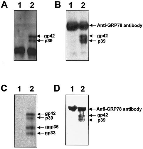
FIG. 4.
GRP78/BiP binds to the L protein in vivo. (A) The COS7 cell lysates, transfected with pCDNA3 as a control vector (lane 1) or the L protein expression plasmid, pCDNAL (lane 2), were subjected to Western blotting with the humanized version of anti-pre-S1 antibody. (B) The COS7 cell lysates, transfected with pCDNA3 (lane 1) or pCDNAL (lane 2), were immunoprecipitated with anti-BiP antibody. The immunoprecipitates were subjected to Western blot analysis using the humanized version of anti-pre-S1 antibody and HRP-conjugated anti-human IgG antibody. (C) The HepG2 cell lysates, transfected with mock DNA (lane 1) or pHBV5.2 (lane 2), were subjected to Western blotting with anti-pre-S2 antibody. (D) The HepG2 cell lysates, transfected with mock DNA (lane 1) or pHBV5.2 (lane 2), were immunoprecipitated with anti-BiP antibody, and the immunoprecipitates were subjected to Western blot analysis using anti-pre-S2 monoclonal antibody and HRP-conjugated anti-murine IgG antibody. The L and M proteins and the heavy chain of anti-BiP antibody that was reacted with HRP-conjugated anti-murine IgG antibody or anti-human IgG antibody are indicated. Molecular size markers (in kilodaltons) (lane M) are shown on the right.

