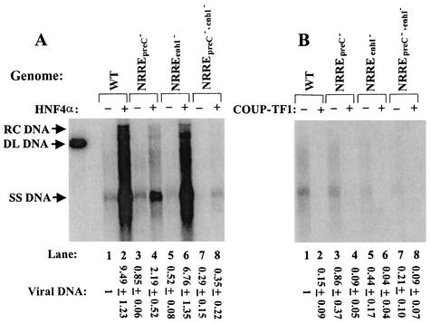
FIG. 3.
Activation and repression of synthesis of viral DNA by HNF4α (A) and COUP-TF1 (B), respectively. Shown are autoradiograms of Southern blot analysis of viral DNA isolated from nucleocapsids present in the cytoplasm of Huh7 cells cotransfected with the indicated HBV genome and 2 μg of pCDMHNF4 (A) or 1 μg of pRSVCOUP-TF1 (B). One-third of the DNA from a 60-mm-diameter dish of cells was loaded in each lane. The total amount of viral DNA in each lane, from relaxed circular (RC) through single-stranded (SS) DNA, was quantified with a PhosphorImager. The smears between the specifically indicated DNA structures are the result of HBV DNA with incomplete, heterogeneous-length plus strands. Numbers at the bottom are means ± standard errors of the data relative to the wild type (WT) obtained from three experiments similar to the one for which results are shown. DL, duplex linear.

