
FIG. 7.
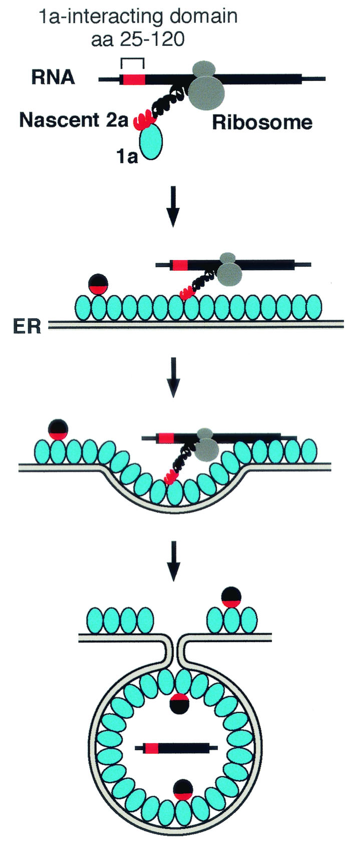
Model for the 2a ORF-dependent 1a responsiveness of highly translated 2a mRNAs. The successive panels illustrate initiation of interaction between 1a and the N terminus of the nascent 2a peptide still associated with its mRNA through the ribosome, localization of the translating 2a peptide and RNA to the ER membrane by 1a, and sequestration of 2a and the RNA into the capsid-like, membrane-bound spherules formed by multiple 1a proteins (37). The red and black spheres represent mature 2a protein, with the N-proximal 1a-interacting domain of 2a indicated by red as in the nascent 2a protein.