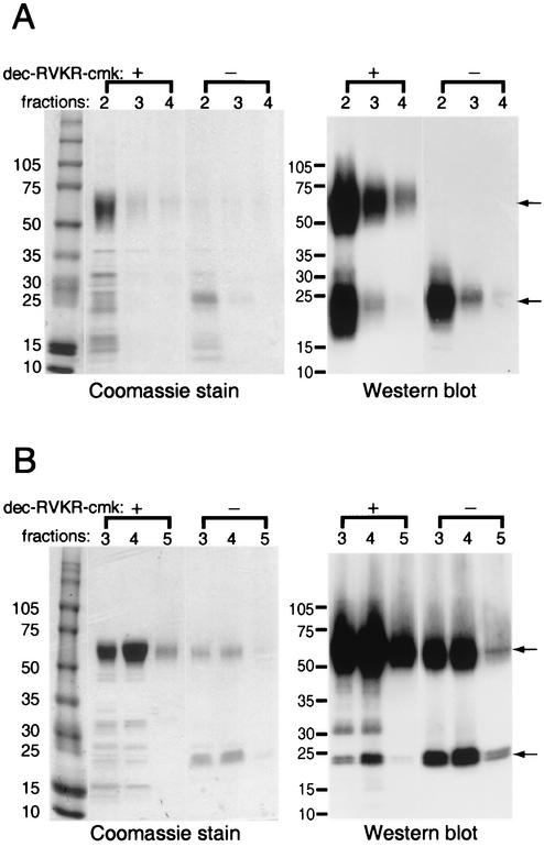
FIG. 3.
Expression of recombinant MC54L protein in the presence (+) or absence (−) of a furin inhibitor. (A) 293T cells were transiently transfected with a plasmid encoding MC54L protein with a C-terminal six-histidine tag. (B) BS-C-1 cells were infected with a recombinant vaccinia virus encoding MC54L protein with a C-terminal six-histidine tag. Transfected or infected cells were incubated in medium with (+) or without (−) 50 μM dec-RVKR-cmk. Secreted recombinant MC54L proteins were purified by metal affinity chromatography, and the eluted fractions indicated by number were subjected to SDS-PAGE. The proteins were detected by Coomassie blue staining (left) or by chemiluminescence after Western blotting with a MAb to the polyhistidine tag (right). The arrows point to the full-length (top) and C-terminal fragment (bottom) forms of the MC54L protein. The values on the left indicate the mobilities and masses in kilodaltons of marker proteins.

