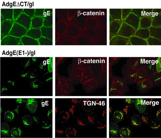
FIG. 2.
Confocal immunofluorescence microscopy of cells expressing gEΔCT/gI. HaCaT cells were infected with either AdgEΔCT (150 PFU/cell), AdtetgI (150 PFU/cell) and AdTet-trans (80 PFU/cell), or AdgE(E1−) and AdgI(E1−) with 150 PFU of each per cell. After 20 to 24 h the cells were fixed, permeabilized, and stained for gE with MAb 3114 and simultaneously with anti-β-catenin or anti-TGN46 antibodies.

