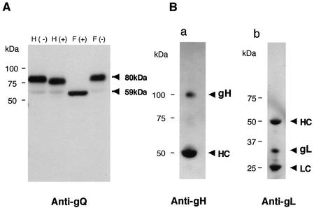
FIG. 7.
Immunoblotting of purified HHV-6A virions. (A) Purified virions were lysed in TNE buffer (10 mM Tris-HCl [pH 7.8], 1% Nonidet P-40, 0.15 M NaCl, 1 mM EDTA), digested with endo H (lanes H) or PNGase F (lanes F), and resolved by SDS-PAGE, and the gels were electrotransferred to PVDF membranes. The blots were probed with the anti-gQ MAb, AU100-119. (B) Lysates of purified virions were immunoprecipitated with AU100-119, electroblotted, and probed with anti-gH antiserum (a) or anti-gL antiserum (b). HC, immunoglobulin heavy chain of the immunoprecipitating antibody. LC, immunoglobulin light chain of the immunoprecipitating antibody. Numbers beside the panels show molecular masses in kilodaltons.

