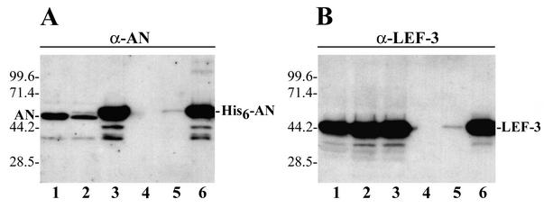
FIG. 2.
Western blot analysis of the binding of AN and LEF-3 to a Ni-NTA resin. Equal portions (3 μl) from the crude extracts and the samples collected from Ni-NTA columns were analyzed by SDS-10% PAGE followed by Western blot analysis with polyclonal antibodies to AN (A) or LEF-3 (B). The extracts (5 ml) from 0.9 × 10⁸ Sf9 cells infected (multiplicity of ∼4) with wt AcMNPV (48 h p.i.), vAcHISAN (18 h p.i.), or vAcHISAN (48 h p.i.) were analyzed in lanes 1, 2, and 3, respectively. The extracts were clarified by centrifugation and processed on identical Ni-NTA columns (0.6 ml), as described in Materials and Methods. The bound proteins were eluted with 3 ml of buffer C containing 150 mM imidazole, and the samples obtained from the cells infected with wt AcMNPV (48 h p.i.), vAcHISAN (18 h p.i.), or vAcHISAN (48 h p.i.) were analyzed in lanes 4, 5, and 6, respectively. The molecular masses of protein markers (in kilodaltons) are shown on the left sides of the panels.

