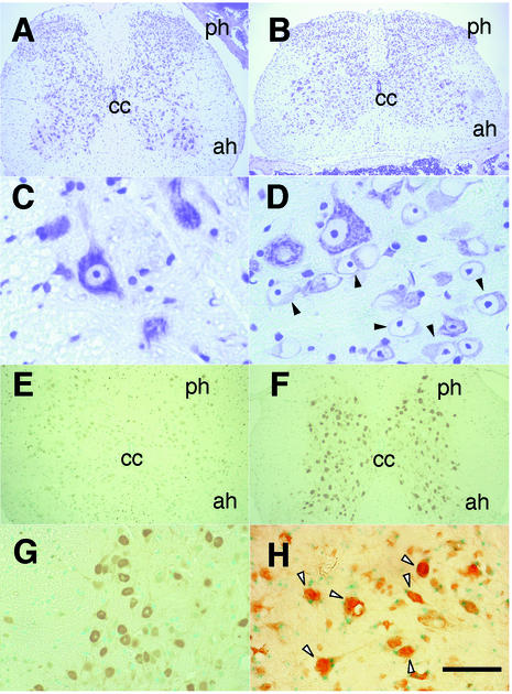
FIG. 3.
Histopathological changes in CNS neurons of maf mutant mice. (A to D) Nissl staining of spinal cord sections from 10-week-old mafG+/−::mafK+/− control mice (A and C) and mafG−/−::mafK+/− mutant mice (B and D) displaying myoclonus. Solid arrowheads (D), swollen nuclei with minimal cytoplasm in the affected mice. (E to G) Immunostaining for ubiquitin in spinal cord sections from control mafG+/−::mafK+/− mice (E) and mafG−/−::mafK+/− mutant mice (F and G). The mutants display far more intense staining, primarily in neuronal nuclei (G). (H) Double staining for β-galactosidase activity and ubiquitin immunohistological activity on brain stem sections prepared from 4-week-old mafG−/−::mafK+/− mutant mice. Note that all the cells in which ubiquitin has accumulated (intense brown signal) are also LacZ positive (open arrowheads). Scale bar, 400 (A, B, E, and F), 40 (C and D), 120 (G), or 60 μm (H). Abbreviations are as defined in the legend to Fig. 2.

