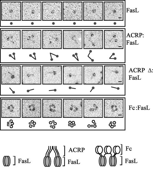
FIG. 2.
Rotary shadowing electron microscopy of recombinant FasL. Six representative images of FasL, ACRP:FasL, ACRPΔ:FasL, and Fc:FasL preparations are shown, with a schematized interpretation of the picture. Filled circles, trimeric FasL; black rods, collagen domain of ACRP or ACRPΔ; open circles, dimeric Fc portion of IgG1; thin lines, possible connectivity between FasL and Fc domains. For Fc:FasL, alternative interpretations are possible regarding the identification of FasL and Fc domains and regarding the connectivity between domains. Pictures were taken at a magnification of ×150,000, and a scale of 10 nm is indicated (bars). Schematic representations of the hexameric forms of ACRP:FasL and Fc:FasL are shown at the bottom of the figure.

