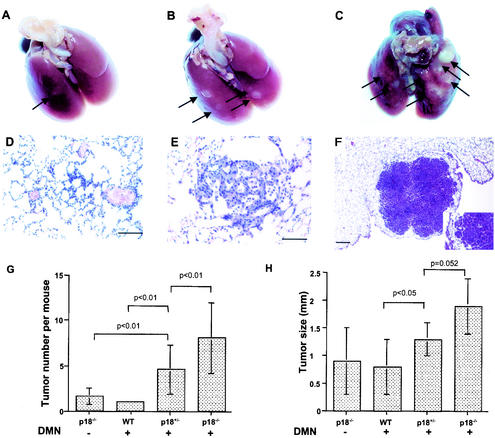
FIG. 3.
Mutation in p18 gene sensitized mice to DMN-induced lung tumors. (A to C) The rate of lung tumor development varied between age-matched (11 months) p18+/+, p18+/−, and p18−/− mice after DMN exposure. Gross alveolar and bronchiolar adenomas (white spots) are identified by arrows. Note that p18 mutant lungs developed more tumors. (D to F) Alveolar or bronchiolar origin of lung tumor development. Lungs of DMN-treated mice at different ages were microscopically examined after hematoxylin-eaosin staining and exhibited alveolar or bronchiolar hyperplasia (D), papillary alveolar or bronchiolar adenoma with mucinous cell differentiation (E), and alveolar or bronchiolar carcinoma (F). The inset in panel F is a higher magnification showing tumor infiltration of adjacent pulmonary parenchyma. (G) The number of lung tumors that developed in each mouse exposed to DMN increases with p18 gene mutation. A statistically significant difference in tumor multiplicity was seen in mice of a different p18 genotype following exposure to DMN. WT, wild type. (H) The average size of DMN-induced lung tumors was larger in p18 mutant mice than in wild-type mice.

