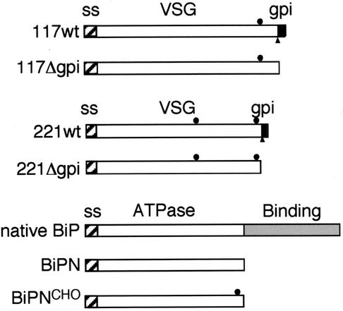
FIG. 1.
Diagram of secretory reporters. Diagrammatic representations of VSG 117, VSG 221, and BiP reporters are shown (scale approximate.) The hatched boxes denote N-terminal signal sequences. The filled circles indicate N-linked glycans. The black boxes signify the GPI attachment peptide, and the filled triangle shows the site of cleavage and GPI attachment to VSG. The native BiP structure is shown for comparison. The BiP ATPase and peptide binding domains are indicated.

