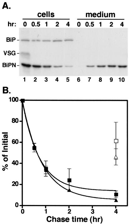
FIG. 2.
Secretion of BiPN. (A) Bloodstream 221 cells expressing transgenic BiPN reporter were pulse-radiolabeled for 10 min with 35S-labeled Met-Cys and then chased for 4 h. BiP polypeptides were immunoprecipitated from cell and medium fractions at the indicated times. Samples were then analyzed by sodium dodecyl sulfate-polyacrylamide gel electrophoresis (SDS-PAGE) and fluorography. Each lane has 5 × 10⁶ cell equivalents. The positions of endogenous BiP, VSG, and the BiPN reporter have been indicated. (B) The experiment shown in panel A was performed in triplicate, and the amount of both the cell-associated and secreted forms of the two different BiPN reporters were quantified by phosphorimaging. Cell-associated (solid symbols) and medium-accumulated (open symbols; only the final datum point is shown) BiPN as a percentage of time zero (squares, means ± the standard error) are plotted as a function of chase time. The same analysis was also performed on a glycosylated version of BiPN (BiPNCHO, triangles).

