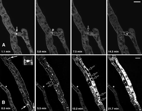
FIG. 2.
A time course of compatible and incompatible hyphal fusion in N. crassa. The figure shows confocal images showing different stages of the hyphal fusion process at the times indicated after staining with the membrane-selective fluorescent dye FM4-64 (30). FM4-64 initially stains the plasma membrane and subsequently becomes visible in internal membranes (23). (A) Compatible hyphal fusion between two N. crassa strains that have identical specificity at all het loci. Initially, two Spitzenkörper (solid arrow; 1.1 min) are present on either side of the region where pore formation will subsequently occur (p; 5.8 min). A persistent ring of fluorescence (open arrow; 7.5 min) is present around the fusion pore after it has formed. Cytoplasmic flow is associated with movement of organelles through the pore, including nuclei and large vacuoles (v; 14.5 min). Note that the FM4-64 dye has become slightly photobleached in these cells during the time course. Bar = 10 μm. Time-lapse movies of hyphal fusion are available at the Fungal Genetics and Biology website (30) and www.Neurospora.org. (B) Incompatible hyphal fusion between two N. crassa strains that differ in allelic specificity at het-c. Heterokaryotic cells become compartmentalized by occlusion of septa (solid arrows and inset; 0.5 min). Increased cytoplasmic staining is due to the permeabilization of the plasma membrane and nonspecific incorporation of FM4-64 into internal membranes. Compare the incompatible fusion cells with the relatively constant level of staining of nearby healthy hypha (*) (the healthy hypha becomes slightly photobleached over time). Large vacuoles form within incompatible fusion cells (open arrows; 5.5 and 13.2 min) but eventually burst as cell death proceeds (21.7 min). Bar = 10 μm. Images are courtesy of D. J. Jacobson (University of California—Berkeley) and N. D. Read (University of Edinburgh).

