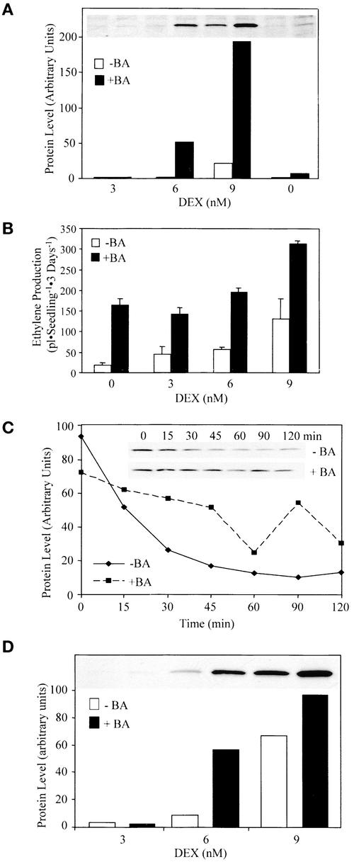
Figure 7.
Cytokinin Increases the Stability of the ACS5 Protein.
(A) Cytokinin causes an increase in the steady state level of myc-ACS5WT. Seedlings harboring the myc-ACS5WT transgene were grown in the dark for 3 days on MS medium containing the indicated amount of DEX in the presence of 5 μM benzyladenine (+BA) or a DMSO vehicle control (−BA) as indicated. Proteins were extracted from the seedlings and analyzed by immunoblotting using an anti-myc monoclonal antibody. The inset shows an image of the original film, and the graph is a depiction of the quantification of each signal. The lanes in the blot correspond to those indicated in the graph.
(B) Measurement of ethylene production from a myc-ACS5WT transgenic line. Seedlings were grown on MS medium containing the indicated amount of DEX for 3 days in the dark in the presence (closed bars) or absence (open bars) of 5 μM benzyladenine in capped GC vials, and the level of ethylene accumulated was measured using GC analysis.
(C) Cytokinin causes an increase in the half-life of ACS5. Myc-ACS5WT transgenic seedlings were grown for 3 days in the dark on MS plates containing 5 μM benzyladenine plus 15 nM DEX or a DMSO vehicle control plus 20 nM DEX. The seedlings were washed in liquid MS medium lacking DEX and then suspended in liquid MS medium lacking DEX plus either benzyladenine or DMSO and containing the protein synthesis inhibitor cycloheximide at time 0. At various times (indicated in minutes above each lane), the seedlings were harvested, and protein extracts were analyzed by immunoblotting using an anti-myc monoclonal antibody probe. The inset shows an image of the immunoblot that was quantified and the level of protein plotted versus time after the inhibition of protein synthesis.
(D) Cytokinin causes an increase in the steady state level of myc-ACS5eto2. Transgenic myc-ACS5eto2 seedlings were grown for 3 days in the dark on MS medium supplemented with 5 μM benzyladenine or a DMSO vehicle control and various concentrations of DEX as indicated. The proteins were extracted and analyzed by immunoblotting using an anti-myc monoclonal antibody probe. The inset shows an image of the protein blot that was quantified.

