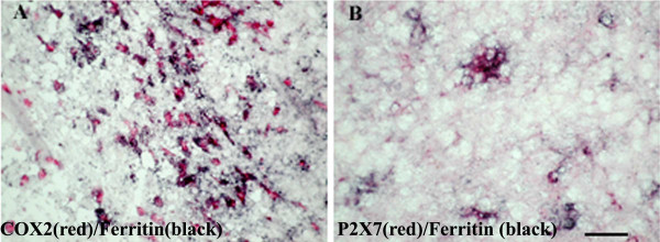
Figure 10.
Co-localisation studies of COX-2 and P2X7 in MS spinal cord. Double staining of COX-2 or P2X7 cells with the microglia marker ferritin in MS spinal cord. Sections were incubated with mixture of A) Ferritin (black) and COX-2 (red) antibodies or B) ferritin (black) and P2X7 (red). The majority of COX-2 or P2X7 immunoreactive cells appear to be microglial cells/macrophages. Scale bar = 100 μm.

