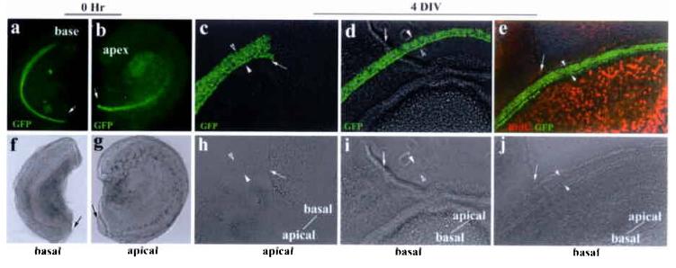
Fig. 2.

Unidirectional extension of the organ of Corti in vitro. a-b: bisected basal (a) and apical (b) portions of the same cochlear duct. Nuclei-localized GFP is expressed under the control of Math1 enhancers4, marking the hair cells in live cultures. The bisection sites were indicated by arrows. Both the basal (basal culture) and apical (apical culture) portions of the cochlea were used for in vitro cultures (c-e). Note that the direction of prospective extensions beyond the wound sites for the basal or the apical cultures will be basal-to-apical or apical-to-basal, respectively. c-e: both the basal (basal culture) and apical (apical culture) portions of the same cochlea (a-b) were cultured and extensions were monitored using the cutting wound sites (indicated by arrows) as reference points (c-e). Hair cells (GFP+) differentiated normally in the apical culture after 4 DIV (c). However, the rows of hair cells did not pass the bisection site (c), indicating no apical-to-basal extension in the apical culture. The rows of hair cells in the basal cultures (d and e) extended past the bisection sites (d, e) from basal-to-apical direction. No BrdU incorporation in the organ of Corti was detected in extended basal cultures (e). The rows of hair cells were bracketed with a pair of arrowheads, and the orientation of the explants was indicated in (c-e).The bottom panels (f-j) are corresponding phase images of the upper panels.
