
Fig. 3.
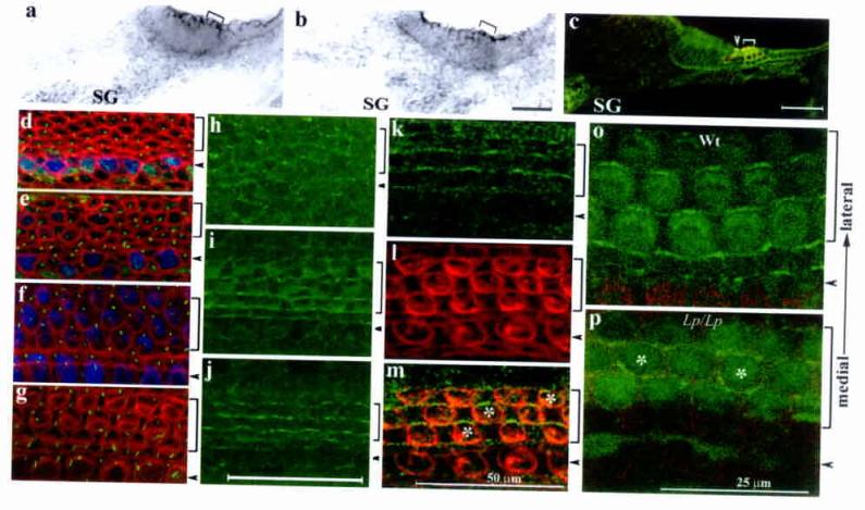
Presence of a functional PCP pathway in the developing organ of Corti. a-c: in situ hybridization of Celsr1 (a) and Dvl1 (b) at E16.5, and DVL2-EGFP signals (c) at E18.5. The brackets in (a-b) indicate the organ of Corti. d-g: Confocal scans of the E17.5 organ of Corti at 80% (d), 75% (e), 50% (f), and 25% (g) from the base of the cochlear duct. At the base (g), hair cells are differentiated and stereocilia (red) are visible. In the medial region (f), the stereocilia are less developed and not visible (red). However, both inner and outer hair cells are differentiated (blue) and the kinocilia (green) are eccentric, indicating a polarized region. At the apical region (d-e), myosinVI expression (blue) has yet to complete in the outer hair cell region and the kinocilia (green) are centric, indicating a transition stage prior to the polarization process. h-m: Confocal scanning of DVL2-EGFP (green) in a whole mount organ of Corti at E17.5 from the corresponding apical (h), medial (i), and basal (j) regions as shown in d-g. Counter-staining with hair cell membrane (l, red) and DVL2-EGFP (k) at E18.5 indicated DVL2-EGFP signal at the lateral side of hair cells (m) o-p: DVL2-EGFP in wild-type (o) and Lp/Lp (p) littermates at E18.5. Note the loss of the wild-type (o) subcellular localization of DVL2-EGFP in the Lp/Lp mutant (p, the asterisks mark hair cells with DVL2-EGFP at the medial side). Arrowheads and brackets (c-p) mark the inner and outer hair cells, respectively. Scale: 50 μm (a-m).