
Fig. 6.
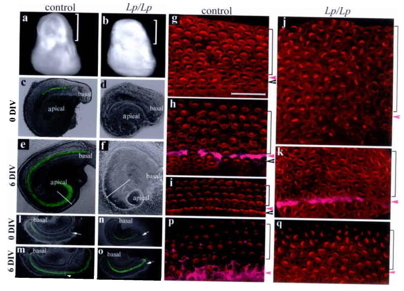
Direct requirement of PCP pathway in extension and planar cell polarity of the organ of Corti. a-b: inner ears from control and Lp/Lp littermates at E14.5. Brackets mark the cochlear portion of the inner ear. c-f: intact cochlear ducts from control (c and e) and Lp/Lp (d and f) cultured 6 DIV. Diameters of the cochlear ducts at the comparable apical regions were outlined (e and f). Hair cells were visualized by Math1-EGFP. g-k: confocal images stereocilia (phalloidin) for control (g-i) and Lp/Lp (j-k) cultures at the apical (g and j), medial (h and k), and basal (i) regions. Scale: 25 μm. l-q: bisected basal regions of the cochlear ducts from wild type (l-m) and Lp/Lp (n-o) cultured for 6 DIV, and the confocal images of stereocilia near the bisection sites for the wildtype (p) and Lp/Lp (q). Arrows in l-o indicate the bisection sites. Arrowheads and brackets in (g-k, p-q) mark the inner and outer hair cell regions, respectively, and magenta arrowheads mark the pillar cell region that was stained with p75 (h, k, p,magenta).