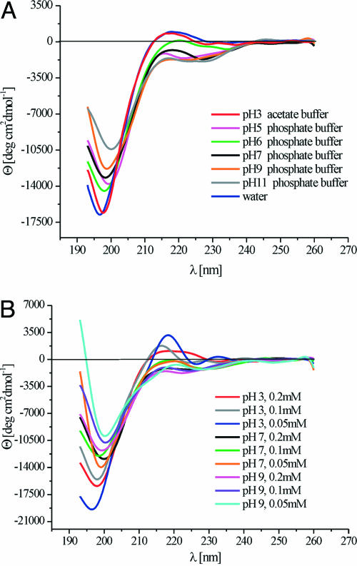
Fig. 1.
CD spectra of the XAO peptide. (A) XAO peptide at 0.2 mM in 0.2 M acetate buffer at pH = 3 and 5°C, 0.2 mM in water at 5°C, and in phosphate buffers (0.007 M–0.25 M) at pH = 5, 6, 7, 9, and 11 at 5°C. (B) XAO peptide in 0.2 M acetate buffer at pH = 3 with peptide concentrations of 0.2 mM, 0.1 mM, and 0.05 mM and in phosphate buffers (0.01 M and 0.15 M) at pH = 7 and pH = 9 with peptide concentrations of 0.2 mM, 0.1 mM, and 0.05 mM at 1°C.

