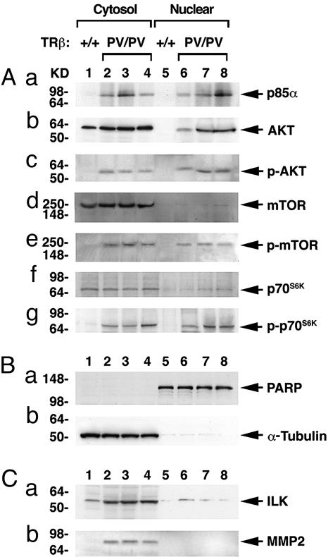
Fig. 5.
Activation of the PI3K–AKT–p70S6K and ILK–MMP2 pathwaysin the thyroid of TRβPV/PV mice. Pooled extracts from thyroids of 12 wild-type mice or 3 TRβPV/PV mice were separated into nuclear and cytosolic fractions, as described in Materials and Methods. Western blot analysis was carried out as described in Materials and Methods to determine cytosolic and nuclear abundance of the following proteins: p85α (Aa), AKT (Ab), p-AKT(S473) (Ac), mTOR (Ad), p-mTOR (Ae), p70S6K (Af), and p-p70S6K (Ag). (B) The expression of PARP (a) or α-tubulin (b) was used for monitoring the quality of nuclear and cytosolic fractions as well as for loading controls for the Western blot analysis shown in A and C. (C) Increased protein abundance of ILK (a) and MMP2 (b) in the cytosolic fraction of thyroid tumors of TRβPV/PV mice.

