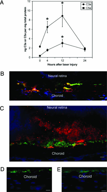
Fig. 3.
Laser injury-induced complement in wild-type mice. ELISA demonstrated increased levels of C3a and C5a in the RPE/choroid within 4 h of injury and a maximum at 12 h after injury. (A) ∗, P < 0.05 compared with PBS. C3 (B) and C5 (C) were deposited (red) in the proximity of RPE cells (green) in the area of injury 4 h after injury. Areas of complement colocalization with RPE cells appear yellow. No deposition of C3 (D) or C5 (E) was identified in unlasered areas. (Scale bars, 5 μm.) DAPI staining appears blue.

