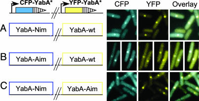
Fig. 5.
Restoration of YabA-Nim localization byYabA-Aim. YabA-Nim and YabA-Aim proteins fused to CFP (blue) were coexpressed with a wild-type YabA fused to YFP (yellow) (A and B, respectively). YabA-Nim and YabA-Aim proteins fused to CFP (blue) or YFP (yellow), respectively, were also coexpressed in the same cells (C). Because the expression of the CFP-fusions was inducible by xylose, the strains were grown on plates containing a low amount (0–0.1%) of xylose to avoid quenching of the YFP signal by the CFP derivative. Using cells with YFP- or CFP-YabA foci, we verified that there was no detectable level of fluorescence crossover of YFP into the CFP channel and vice versa (data not shown).

